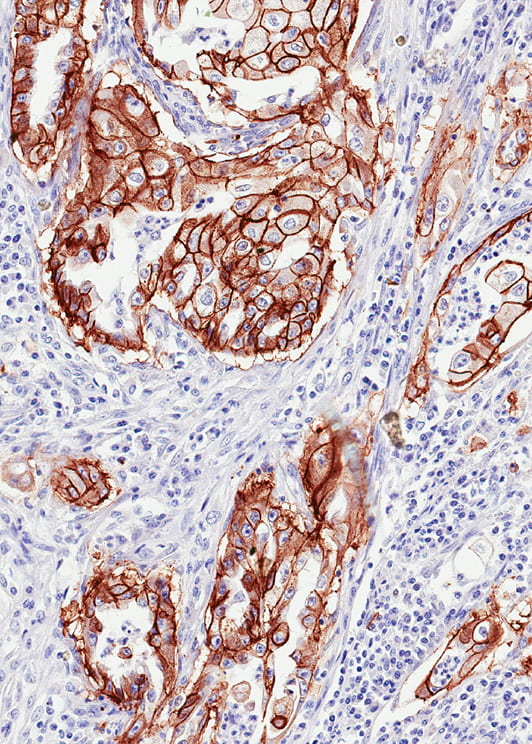
Comprehensive studies of disease pathogenesis in vivo are impossible without comparative pathology. Our work in this area includes development of new pathological classifications allowing direct comparison of existing human and veterinary classifications, development of didactic materials compensating for the current lack of formal training of comparative mouse and human pathologists, and teaching at workshops serving to the same purpose (Bolon et al., 2011; Ince et al., 2008; Kaufman et al., 2010; Nikitin et al., 2004; Sundberg et al., 2018; Valli et al., 2007)
Our work in the area of comparative pathology provides essential ground for development of stem cell pathology. We define stem cell pathology as an area of pathology, which focuses on studying the roles of stem cells in disease pathogenesis, identifies pathological consequences of stem cell transplantation, and evaluates side effects of genetic and epigenetic manipulations of stem cells (Fu et al, 2018). The goals of stem cell pathology are to evaluate consequences of genetic manipulations in stem cells, study in situ contributions of stem cells to normal and pathological regeneration, and to assess organismal reactions to transplantations of various bioengineering constructs. As pertinent to our own research, stem cell pathology supports interpretation of cell lineage hierarchies identified by single-cell transcriptomic and cell lineage tracing approaches. Stem cell pathology also offers new perspective to our cross-disciplinary collaborations in technology-oriented areas, such as biomedical engineering, nonlinear microscopy, intravital imaging and nanotechnology (Chandler et al, 2012; Choi et al., 2007; Flesken-Nikitin et al., 2007, Lu et al., 2017; Lu et al., 2019, Williams et al., 2010; Zipfel et al. 2003).
To facilitate introduction to the mouse pathology, we are offering graduate course VTBMS/TOX 7010 “Mouse and stem cell pathology”. This course open to all graduate students, postdocs and faculty at Cornell.
Since 2002, we are offering the Practical Workshop Series on the Pathology of Mouse Models for Human Disease held in the Jackson Laboratory every fall (Sundberg et al., 2018, 2017). This course is designed for participants with medical background. Minimum qualification of M.D. or D.V.M. (or equivalent) is required.
Select publications
- Lu, Y.-C., Chu, T., Hall, M. S., Fu, D. J., Shi, Q., Chiu, A., An, D., Wang, L., Pardo, Y., Southard, T., Danko, C., Liphardt, J., Nikitin, A. Yu., Wu, M., Fischbach, C., Coonrod, S., and Ma, M. (2019). Physical confinement induces malignant transformation in mammary epithelial cells. Biomaterials 217:119307. doi.org/10.1016/j.biomaterials.2019.119307. PMID: 31271857
- Fu, D.-J., Miller, A. D., Southard, T. L., Flesken-Nikitin, A., Ellenson, L. H., and Nikitin, A. Yu. (2018). Stem cell pathology. Annual Review of Pathology: Mechanisms of Disease. 13:4.1–4.22. DOI: 10.1146/annurev-pathol-020117-043935. PMID: 29059010.
- Sundberg, J. P., Boyd, K., HogenEsch, H., Nikitin, A. Yu., Treuting, P. M., and Ward, J. M. (2018). Training Mouse Pathologists: 16th annual workshop on the pathology of mouse models of human disease. Lab Animal. 47:38-40. doi: 10.1038/laban.1399. PMID: 29384517 PMCID: PMC5857948.
- Lu, Y.-C., Fu, D.-J., An, D., Chiu, A., Schwartz, R., Nikitin, A. Yu., and Ma, M. (2017). Scalable production and cryo-storage of organoids using core-shell decoupled hydrogel capsules. Adv. Biosys. 1700165 doi: 10.1002/adbi.201700165. PMID: 29607405 PMCID: PMC5870136.
- Chandler, E. M., Seo, B. R., Califano, J. P., Andresen Eguiluz, R. C., Lee, J. S., Yoon, C. J., Tims, D. T., Wang, J. X., Cheng, L., Mohanan, S., Buckley, M. R., Cohen, I., Nikitin, A. Yu., Williams, R. M., Gourdon, D., Reinhart-King, C. A., and Fischbach, C. (2012). Implanted adipose progenitor cells as physicochemical regulators of breast cancer. Proc. Natl. Acad. Sci. USA, 109:9786-9791.
- Bolon, B., Altrock, B., Barthold, S., Baumgarth, N., Besselsen, D., Boivin, G., Boyd, K., Brayton, C., Cardiff, R., Couto, S, Eaton, K., Foreman, O., Griffey, S., La Perle, K., Lairmore, M., Liu, C., Meyerholz. D., Nikitin, A. Yu., Schoeb, T., Schwahn, D., Sellers, R., Sundberg, J., Tolwani, R., Valli, V., and Zink, M. C. (2011). NIH plan will hinder translational studies. Nature 47: 36–37.
- Bolon, B., Altrock, B., Barthold, S., Baumgarth, N., Besselsen, D., Boivin, G., Boyd, K., Brayton, C., Cardiff, R., Couto, S, Eaton, K., Foreman, O., Griffey, S., La Perle, K., Lairmore, M., Liu, C., Meyerholz. D., Nikitin, A. Yu., Schoeb, T., Schwahn, D., Sellers, R., Sundberg, J., Tolwani, R., Valli, V., and Zink, M. C. (2011). Advancing translational research. Science 331: 1516-1517.
- Williams, R. M., Flesken-Nikitin, A., Ellenson, L. H., Connolly, D. C., Hamilton, T. C., Nikitin, A. Yu., and Zipfel, W. R. (2010). Strategies for high-resolution imaging of epithelial ovarian cancer by laparoscopic nonlinear microscopy. Translational Oncology. 3:181-194 . PMID: 20563260, PMCID: PMC2887648.
- Kaufman M. H., Nikitin, A. Yu., and Sundberg, J. P. (2010). Histologic Basis of Mouse Endocrine System Development: A Comparative Analysis. CRC Press. Boca Raton, FL., 232 p.
- Ince, T. A., Ward, J. M., Valli, V. E., Sgroi, D., Nikitin, A. Yu., Loda, M. F., Griffey, S. M., Crum, C. P., Crawford, J. M., Bronson, R. T., Brayton, C., and Cardiff, R. D. (2008). “Do-it-yourself (DIY) Pathology”. Nature Biotechnology. 26:978-979.
- Valli, T., Barthold, S. W., Ward, J. E., Brayton, C., Nikitin, A. Yu., Borowsky, A. D., Bronson, R. T., Cardiff, R. D., Sundberg, J., and Ince, T. (2007). Over 60% of NIH extramural funding involves animal-related research. Vet. Pathology 44: 962-963.
- Flesken-Nikitin, A., Toshkov, I.A., Nascar, J., Tyner, K. M., Williams, R. M., Zipfel, W. R., Giannelis, E., and Nikitin, A. Yu. (2007). Toxicity and biomedical imaging of layered nanohybrids in the mouse. Tox. Pathology 35: 804-810.
- Choi, J., Burns, A. A., Williams, R. M., Zhou, Z., Flesken-Nikitin, A., Zipfel, R. W., Wiesner, U., and Nikitin, A. Yu. (2007). Core-shell silica nanoparticles as fluorescent biological labels for nanomedicine applications. J. Biomed. Optics 12: 064007.
- Nikitin, A. Yu. , Alcaraz, A., Anver, M., Bronson, R. T., Cardiff, R. D., Dixon, D., Fraire, A. E., Gabrielson, E. W., Gunning, W. T., Haines, D. C., Kaufman, M. H., Linnoila, I., Maronpot, R. R., Rabson, A. S., Reddick, R. L., Rehm, S., Rozengurt, N., Schuller, H. M., Shmidt, E. N., Travis, W. D., Ward, J. M., and Jacks, T. (2004). Classification of proliferative pulmonary lesions of the mouse: Recommendations of the Mouse Models of Human Cancers Consortium. Cancer Res., 64: 2307-2316.
- Zipfel, W. R., Williams, R. M., Christie, R., Nikitin, A. Yu., Hyman, B. T., and Webb, W. W. (2003). Live tissue intrinsic emission microscopy using multiphoton-excited fluorescence and second harmonic generation. Proc. Natl. Acad. Sci. USA, 100: 7075-7080.